Digital First Dentistry › Scan, See, Scale: the three moves of digital-first dentistry
Scan, See, Scale: the three moves of digital-first dentistry
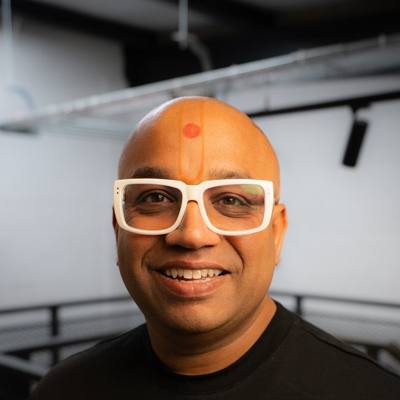 By Dr Bhavin Bhatt BDS MFGDP RCS (Eng) · GDC 76358
Updated November 2026
In the first post in this series I argued that most dentistry still starts when something breaks, and that the real gap is between what the dentist can see and what the patient can understand. The white paper on digital-first dentistry answers that gap with three moves: Scan, See, Scale. They sound simple. The point is the order, because each one only works if the one before it has happened.
Scan: make it part of the check-up
The first move is to make the intraoral scan part of the routine examination, not something saved for patients who have already agreed to a crown or aligners. Most practices with a scanner work the other way round. The scanner comes out only after treatment has been decided, to take the impression. That is what I call digital second: the scanner makes things, but it rarely finds them.
A full-arch scan at the check-up is quick, it does not hurt, and it can be repeated at every visit. Each scan becomes part of the record. That matters later, because the most useful thing a scan can tell you is often not what is there today, but what has changed since last time.
In practice this means deciding a few things in advance: who takes the scan (many practices train nurses to do it), which patients come first (heavily restored mouths are a good start), and where in the appointment it sits, so it is not squeezed in at the end.
See: the same picture on one screen
The second move is the heart of the idea. The scan goes on the screen and the patient looks at their own teeth alongside you. A worn incisal edge, a tooth that has drifted, a cracked cusp, a filling with a broken margin, gum that has receded, a bite that is hitting hard on one side: these stop being things the dentist says and become things the patient can see.
A few habits help. Let the patient look before you speak. Point, then explain in plain words. Where there is an earlier scan, show the two together; a scan comparison turns "I think this has got worse" into something you can both check. And when nothing has changed, say so. Seeing that a tooth is stable is good news, and active monitoring, with a planned recheck, is a proper outcome, not a failure to treat.
Scale: care that follows understanding
The third move is what happens when patients really do see and understand. In the white paper's words, comprehensive care follows demand rather than pressure. Patients who have seen the cracked cusp tend to ask what can be done about it. Some choose to act now, some choose to watch, and both are informed choices.
"Scale" can sound like a sales word, so it is worth being precise. It does not mean doing more treatment on each patient. It means the practice grows because patients trust what they are told, return for reviews, and decide earlier, while the options are smaller: an onlay rather than a crown, a bonded repair rather than a replacement. Informed consent is also stronger when the patient has looked at the problem rather than just heard about it. If you track case acceptance, it is a by-product of understanding, not the aim.
Why the order matters
Each move depends on the one before it. Skip the scan and there is nothing to look at, so you are back to describing problems the patient cannot picture. Scan but skip the seeing, and the scan becomes a recording exercise: data in the software that never reaches the conversation. Jump straight to treatment without either, and you are back to persuasion, which is the part most dentists dislike and most patients can sense.
This is also why buying a scanner is not enough on its own. A scanner used only to make crowns covers part of the first move and none of the second.
What it looks like in an ordinary check-up
- Scan the full arch as part of the examination, before deciding anything.
- Show the scan first and let the patient look.
- Compare with the last scan where there is one.
- Grade each finding simply: stable, to discuss, or at risk.
- Offer options, always including monitoring, and let the patient decide at their own pace.
- Record the scan and the decision, ready for comparison next time.
Can teams make this routine?
Yes, with structure. In a University of Manchester study of 97 UK dentists, currently under peer review, the share of dentists scanning at routine check-ups rose from 10% to 80% within 12 months of structured training. It was a before-and-after evaluation without a control group, so it shows what changed rather than proving why.
If you want to try the three moves this week, the guide to scanning at the routine check-up covers the practical steps, and the check-up planner shows where the minutes can come from in your diary.
This is the second of five posts explaining the white paper in plain English. Next: red, amber, green, and how grading the mouth helps patients understand.
Get the white paper, starter guide and checklist
Free. Enter your email and we will email all three to you.
Check your inbox.
We have sent you a short email to confirm your address. Click the button in it and your free resources arrive straight away. If it is not there in a few minutes, look in your spam or promotions folder.